







This guide explains what a slit lamp examination covers, how its seven illumination techniques work, how to run the exam in a sensible order, and how to choose between digital and portable units.
The slit lamp pairs a low-power microscope with a high-intensity light that narrows to a thin beam, letting the examiner view the eyelids, conjunctiva, cornea, iris and lens in cross-section. Fluorescein dye and dilating drops extend the exam to the tear film and the back of the eye.[2]
What Does a Slit Lamp Test Cover?
The exam looks at every structure at the front of the eye.
Structures examined
- Eyelids and lashes
- Conjunctiva and sclera
- Cornea
- Iris and lens
Dye and drops
Fluorescein, applied with a paper strip or an eyedrop, highlights the cornea and tear layer. Dilating drops take 15 to 20 minutes to work; the exam is then repeated with a handheld lens to view the back of the eye. Patients stay light-sensitive for a few hours afterward.
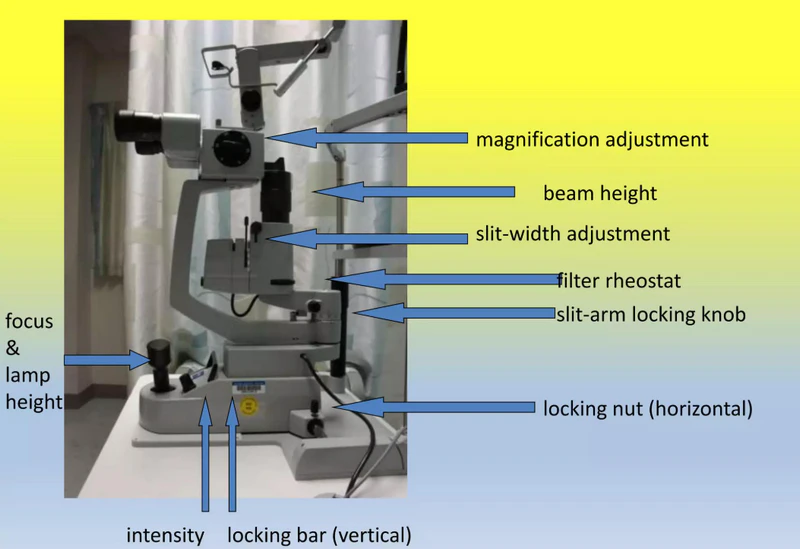
Slit Lamp Parts: Three Systems in One Instrument
Every slit lamp combines three systems:[3]
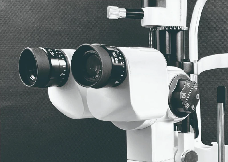
- Observation system: binocular eyepieces and magnification changer
- Illumination system: lamp housing, slit width and height controls, filters
- Mechanical support: chin rest, forehead rest, joystick base
Seven Illumination Techniques
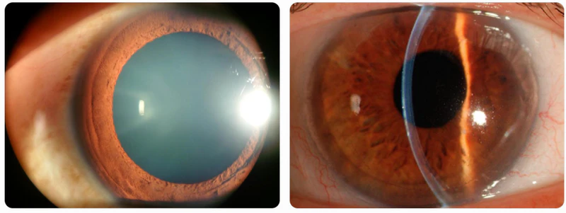
StatPearls lists seven basic techniques, and most exams use at least three.Diffuse illumination uses the widest slit, low to medium magnification and medium to high light intensity.
| Technique | Beam setup | Typical use |
|---|---|---|
| Diffuse | Widest slit, low magnification | Overview of lids, lashes, conjunctiva |
| Direct focal | Narrow to medium slit, oblique beam | Optical section of cornea and lens |
| Indirect | Beam beside the area of interest | Tissue next to a lesion |
| Retroillumination | Light reflected from iris or fundus | Opacities, lens changes |
| Specular reflection | Mirror-like reflex from a surface | Corneal endothelium |
| Sclerotic scatter | Wide beam at the limbus | Corneal haze and edema |
| Oscillating (Koeppe) | Moving beam | Cells and flare in the anterior chamber |
How to Run a Slit Lamp Examination in a Sensible Order
Order matters because bright direct light bothers patients.
Setup
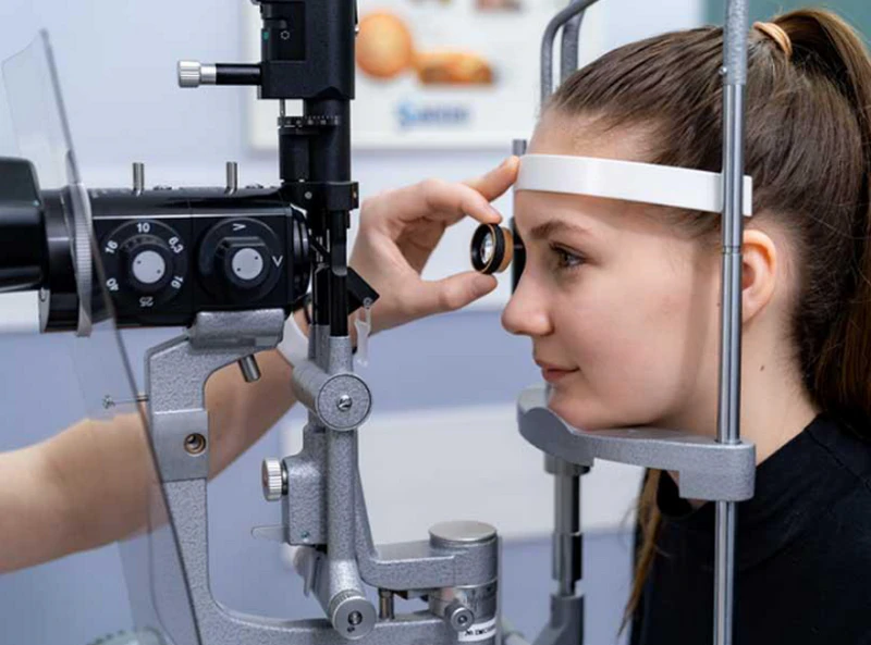
Seat the patient with chin and forehead against the supports, dim the room and keep the observation arm directly in front of the eye.
Sequence
- Start with diffuse illumination at low magnification, with the beam at 30 to 45 degrees to the observation arm
- Move to the corneal layers: epithelium, stroma, endothelium
- Examine the iris, pupil and lens last, since direct illumination is most bothersome there
Intensity
Novices tend to leave brightness at maximum. Turn it down. Most slit lamps offer 10X, 16X and 25X, and some reach 40X.[4]
Housekeeping
Dust on the mirror muddies every view. A small brush, usually included with the accessories, clears it in seconds. Some models also tilt the illumination column toward the patient, up to 20 degrees in 5-degree steps.
Limits: What a Slit Lamp Examination Cannot Show Alone
The slit lamp looks at the front of the eye. To see the retina, the examiner needs dilating drops and a small lens held close to the eye, then repeats the exam. For retinal documentation, a fundus camera or OCT does the job better. Treat the slit lamp test as the first look, not the last.
Slit Lamp Market: Why Equipment Choices Are Changing
The global slit lamps market was USD 311.3 million in 2025 and is forecast to reach USD 468.7 million by 2035, a 4.3% CAGR. Digital slit lamps are forecast to pass USD 127.8 million by 2035 at 4.7%, helped by image capture, telemedicine and EHR integration. ZEISS led with over 21% share in 2025.[1]
Digital or Portable: Which Slit Lamp Fits Your Practice?
Match the instrument to the room, not the catalog.
Digital tabletop
Choose this for a fixed exam lane where records and patient education matter. LINKEYE’s digital slit lamp range includes the SL-200A compact unit, the SL-320 three-step model, and the SL-400 and SL-520 with imaging systems. The SL-520C is AC-powered.
Portable and handheld
- Nursing homes and home visits
- Pediatric and bedridden patients
- Outreach screening
LINKEYE’s portable slit lamp range covers the KJ5S1 handheld unit, the KJ5S3 digital portable microscope and the S150.[6] Both ranges list CE, FDA, ISO 9001 and ISO 13485 certification.[5]
Buyer Checklist Before You Order a Slit Lamp
Before ordering, confirm the following with the supplier.
Optics
- Magnification steps: three-step or five-step, typically 10X to 25X or higher
- Illumination: slit width and height control, filters
- Imaging: built-in capture, if you need records
Paperwork and service
Ask for CE, FDA and ISO 13485 certificates and a written warranty. LINKEYE lists a one-year warranty covering repair or replacement of defective parts.
Best Pick in 2026
For a fixed clinic that needs a documented slit lamp examination, start with a digital tabletop model with an imaging system, such as the SL-520. For mobile care, pair it with a handheld unit like the KJ5S3.
Send us your exam volume and workflow and we will suggest a combination.
FAQs
What is a slit lamp test used for?
A slit lamp test examines the eyelids, conjunctiva, sclera, cornea, iris and lens, and can reveal many eye diseases.
How long does a slit lamp exam take?
A slit lamp exam takes about 5 to 10 minutes for the physical examination of the front and surface structures of the eye.
How long do dilating drops take to work?
It takes 15 to 30 minutes for your eyes to fully dilate after eye drops are administered.
What magnification does a slit lamp use?
Most models offer 10X, 16X and 25X; some extend to 40X.
What is the difference between a digital and a standard slit lamp?
The primary difference is that a digital slit lamp incorporates an integrated camera and software system to capture, store, and display live or recorded images of the eye on a screen, whereas a standard (traditional) slit lamp relies strictly on optical eyepieces for direct observation
References
[1] Global Market Insights. Slit Lamps Market Size and Share, 2026–2035. Accessed October 15, 2026.
[2] MedlinePlus, U.S. National Library of Medicine. Slit-lamp exam. Reviewed January 20, 2025.
[3] StatPearls Publishing. Slit-Lamp Biomicroscope. Accessed October 15, 2026.
[4] Contact Lens Spectrum. 10 Steps to a Better Slit Lamp Examination. November 2006.
[5] Shanghai LINKEYE. Digital Slit Lamp | Precision Eye Examination Tool. Category page, accessed October 15, 2026.
[6] Shanghai LINKEYE. Portable Slit Lamp | Handheld Ophthalmic Slit Lamp Machine. Category page, accessed October 15, 2026.


