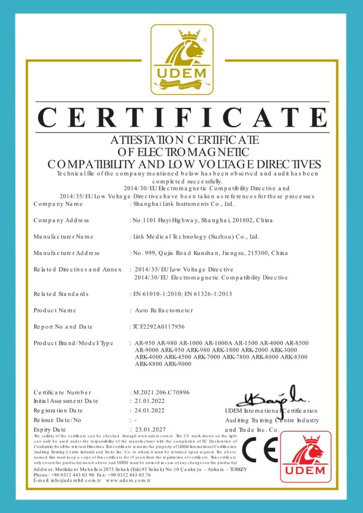


| Parameter | BigVision | 1000T | 1000S | 1000M |
|---|---|---|---|---|
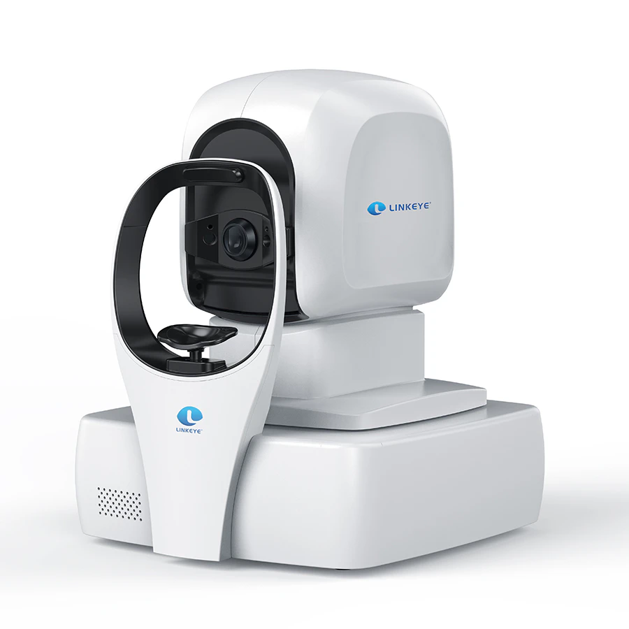
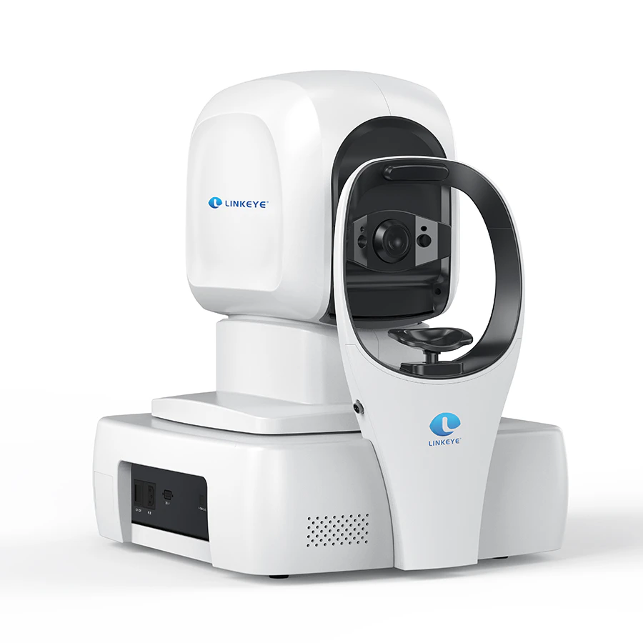
| Jenis | OCT domain spektral (SD-OCT) | OCT domain spektral (SD-OCT) | OCT domain spektral (SD-OCT) | OCT domain spektral (SD-OCT) |
| Energi optik (pada kornea) | ≤750μW | ≤750μW | ≤750μW | ≤750μW |
| Karakteristik pengukuran | Resolusi aksial: 5 μm. Resolusi melintang: 20μm. | Resolusi aksial: 5 μm. Resolusi melintang: 20μm. | Resolusi aksial: 5 μm. Resolusi melintang: 20μm. | Resolusi aksial: 5 μm. Resolusi melintang: 20μm. |
| Karakteristik pemindaian | Kecepatan pemindaian maksimum: ≥ 20.000 kali/detik Kedalaman pemindaian: 2.3mm Jangkauan pemindaian maksimum: 12mm * 9mm | Kecepatan pemindaian A maksimum: ≥ 80.000 kali/detik Kedalaman pemindaian: 2.3mm Jangkauan pemindaian maksimum: 12mm * 9mm | Kecepatan pemindaian A maksimum: ≥ 55000 kali/detik Kedalaman pemindaian: 2,3m Jangkauan pemindaian maksimum: 10mm * 9mm | Kecepatan pemindaian A maksimum: ≥ 55000 kali/detik Kedalaman pemindaian: 2.3mm Jangkauan pemindaian maksimum: 12mm * 9mm |
| Karakteristik sumber cahaya | Panjang gelombang pusat: 840nm Daya optik: ≤ 750μW. Kisaran kompensasi refraktif: -20D~+25D. | Panjang gelombang pusat: 840nm Daya optik: ≤ 750μW. Kisaran kompensasi refraktif: -20D~+25D. | Panjang gelombang pusat: 840nm Daya optik: ≤ 750 μW. Kisaran Kompensasi Bias: -20D~+25D. | Panjang gelombang pusat: 840nm Daya optik: ≤ 750μW. Kisaran Kompensasi Refraksi: -20 D~+25D. |
| Mode pemindaian | Lima mode: Bidang penglihatan yang luas: garis lurus definisi tinggi, enam garis radiasi, area lokal: makula, cakram optik. | Tujuh mode: Bidang penglihatan yang luas: garis lurus definisi tinggi, enam garis radiasi, area: Lokal: makula dan cakram optik. Segmen anterior: garis lurus definisi tinggi, enam garis radiasi. | Tujuh mode: Bidang penglihatan yang luas: garis lurus definisi tinggi, enam garis radiasi, area: Lokal: makula dan cakram optik. Segmen anterior: garis lurus definisi tinggi, enam garis radiasi. | Tujuh mode: Bidang penglihatan yang luas: garis lurus definisi tinggi, enam garis radiasi, area: Lokal: makula dan cakram optik. Segmen anterior: garis lurus definisi tinggi, enam garis radiasi. |
| Rentang pemindaian | 12mm * 9mm Selesaikan akuisisi gambar makula + cakram optik sekaligus. | 12mm * 9mm Selesaikan akuisisi gambar makula + cakram optik sekaligus. | 12mm * 9mm Selesaikan akuisisi gambar makula+cakram optik sekaligus. | 10mm * 9mm Selesaikan akuisisi gambar cakram makula+optik sekaligus. |
| Panjang pemindaian satu baris | 12mm (6mm di segmen anterior) Topografi kornea ruang anterior | 12mm (6mm di segmen anterior) Topografi kornea ruang anterior | 12mm (6mm di segmen anterior) Topografi kornea ruang anterior | 12mm (6mm di segmen anterior) Topografi kornea ruang anterior Topografi kornea |
| Diameter pupil minimum | 2mm | 2mm | 2mm | 2mm |
| Pemosisian otomatis pupil | ✓ | ✓ | ✓ | ✓ |
| Kompensasi otomatis murid | ✓ | ✓ | ✓ | ✓ |
| Gambar fundus | Teknologi pencitraan semua piksel berdasarkan kecepatan pemindaian yang tinggi | Teknologi pencitraan semua piksel berdasarkan kecepatan pemindaian yang tinggi | Teknologi pencitraan semua piksel Berdasarkan kecepatan pemindaian yang tinggi | Teknologi pencitraan semua piksel berdasarkan kecepatan pemindaian yang tinggi |
| Fokus | sepenuhnya otomatis | sepenuhnya otomatis | sepenuhnya otomatis | sepenuhnya otomatis |
| Kecerdasan buatan yang terintegrasi | Membantu diagnosis 25 penyakit mata utama, identifikasi, segmentasi, dan diagnosis yang akurat. | Membantu diagnosis 25 penyakit mata utama, identifikasi, segmentasi, dan diagnosis yang akurat. | Membantu diagnosis 25 penyakit mata utama, identifikasi, segmentasi, dan diagnosis yang akurat. | Membantu diagnosis 25 penyakit mata utama, identifikasi, segmentasi, dan diagnosis yang akurat. |
| Fungsi antarmuka DICOM | ✓ | ✓ | ✓ | ✓ |
| Mode operasi | Pengoperasian sepenuhnya otomatis dan manual | Pengoperasian sepenuhnya otomatis dan manual | Pengoperasian sepenuhnya otomatis dan manual | Pengoperasian sepenuhnya otomatis dan Manual |