






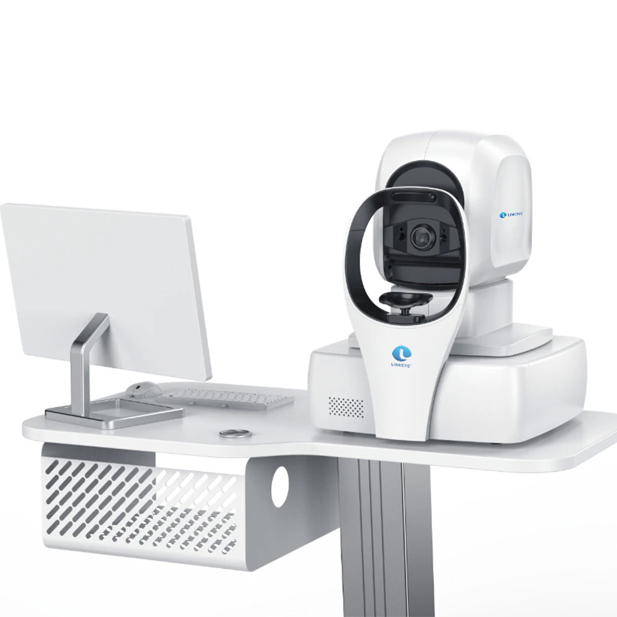
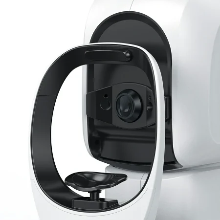
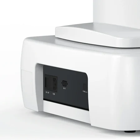

| المعلمة | BigVision | 1000T | 1000S | 1000M |
|---|---|---|---|---|
| النوع | Spectral domain OCT (SD-OCT) | Spectral domain OCT (SD-OCT) | Spectral domain OCT (SD-OCT) | Spectral domain OCT (SD-OCT) |
| Optical energy (at cornea) | ≤750μW | ≤750μW | ≤750μW | ≤750μW |
| Measurement characteristics | Axial resolution: 5 μm. Transverse resolution: 20μm. | Axial resolution: 5 μm. Transverse resolution: 20μm. | Axial resolution: 5 μm. Transverse resolution: 20μm. | Axial resolution: 5 μm. Transverse resolution: 20μm. |
| Scanning characteristics | Maximum a scanning speed: ≥ 20000 times/second Scanning depth: 2.3mm Maximum scanning range: 12mm*9mm | Maximum A scanning speed: ≥ 80,000 times/second Scanning depth: 2.3mm Maximum scanning range: 12mm*9mm | Maximum A scanning speed: ≥ 55000 times/second Scanning depth: 2.3m Maximum scanning range: 10mm*9mm | Maximum A scanning speed: ≥ 55000 times/second Scanning depth: 2.3mm Maximum scanning range: 12mm*9mm |
| Light source characteristics | Central wavelength: 840nm Optical power: ≤ 750μW. Refractive compensation range: -20D~+25D. | Central wavelength: 840nm Optical power: ≤ 750μW. Refractive compensation range: -20D~+25D. | Central wavelength: 840nm Optical power: ≤ 750 μW. Refractive Compensation range: -20D~+25D. | Central wavelength : 840nm Optical power: ≤ 750μW. Refractive Compensation range: -20 D~+25D. |
| Scanning mode | Five modes: Large field of vision: high-definition straight line, six lines of radiation, local area: macula, optic disc. | Seven modes: Large field of vision: high-definition straight line, six lines of radiation, area: Local: macula and optic disc. Anterior segment: high-definition straight line, six lines of radiation. | Seven modes: Large field of vision: high-definition straight line, six lines of radiation, area: Local: macula and optic disc. Anterior segment: high-definition straight line, six lines of radiation. | Seven modes: Large field of vision: high-definition straight line, six lines of radiation, area: Local: macula and optic disc. Anterior segment: high-definition straight line, six lines of radiation. |
| Scanning range | 12mm*9mm Complete the image acquisition of macular+optic disc at one time. | 12mm*9mm Complete the image acquisition of macular+optic disc at one time. | 12mm*9mm Complete the Image acquisition of macular+optic disc at one time. | 10mm*9mm Complete the Image acquisition of macular+opti c disc at one time. |
| Single-line scanning length | 12mm (6mm in anterior segment) Anterior chamber Corneal topography | 12mm (6mm in anterior segment) Anterior chamber Corneal topography | 12mm (6mm In anterior segment) Anterior chamber Corneal topography | 12mm (6mm In anterior segment) Anterio r chamber Corneal topography |
| Minimum pupil diameter | 2mm | 2mm | 2mm | 2mm |
| Pupil automatic positioning | ✓ | ✓ | ✓ | ✓ |
| Pupil automatic compensation | ✓ | ✓ | ✓ | ✓ |
| Fundus image | All-pixel imaging technology based on high scanning speed | All-pixel imaging technology based on high scanning speed | All-pixel imaging technology Based on high scanning speed | All-pixel imaging technology based on high scanning speed |
| Focusing | fully automatic | fully automatic | fully automatic | fully automatic |
| Integrated artificial intelligence | Assisted diagnosis of 25 major ophthalmic diseases, accurate identification, segmentation and diagnosis. | Assisted diagnosis of 25 major ophthalmic diseases, accurate identification, segmentation and diagnosis. | Assisted diagnosis of 25 major ophthalmic diseases, accurate identification, segmentation and diagnosis. | Assisted diagnosis of 25 major ophthalmic diseases, accurate identification, segmentation and diagnosis. |
| DICOM interface function | ✓ | ✓ | ✓ | ✓ |
| Mode of operation | Fully automatic and manual operation | Fully automatic and manual operation | Fully automatic and manual operation | Fully automatic and Manual operation |